facs buffer flow cytometry
The terms flow cytometry and fluorescence-activated cell sorting FACS are often used interchangeably. 2 mL if staining in flow tubes 13.

We Offer Anti Mouse Cd3 Antibody Biot Conjugated Keep As Concentrated Solution Store At 4 C And Protected From Prolonged Exposure To Life Science Index Biot
This Flow Cytometry Staining Buffer is a buffered saline solution.
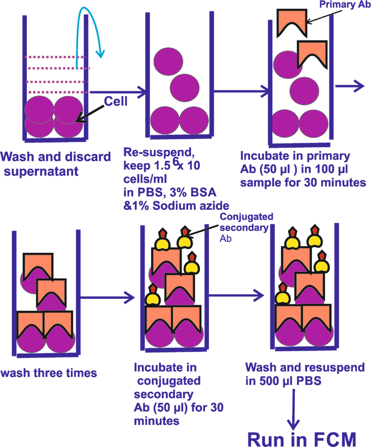
. Primary Antibody Staining 1. 1 Phosphate Buffer Saline or Hanks Buffer CaMg2 free 1 mM EDTA 25 mM HEPES pH 70 05-2 Fetal Bovine Serum Heat-inactivated or 1 BSA 02 μm sterile filtered. Wash cells with FACS buffer a.
Harvest cells and aliquot up to 1 x 10 6 cells100 μL into FACS tubes. Learn More About Spectral Flow Cytometry Fundamentals From Thermo Fisher. Flow Cytometry Scripps Research 10550 North Torrey Pines Road IMM-20 La Jolla CA 92037 tel.
Absence of these ions reduces cation-dependent cell to cell adhesion and prevents clumping. Ad Learn More About Spectral Flow Cytometry Fundamentals From Thermo Fisher Scientific. Learn More About Spectral Flow Cytometry Fundamentals From Thermo Fisher.
They are often described as being displayed in a. 1 mL if staining in Eppendorf tubes b. Ad Products empowering scientists at every stage helping to deliver scientific breakthroughs.
Basic Sorting Buffer 1 x Phosphate Buffered Saline PBS or Hanks Balanced Salt Solution HBSS. Use of FCS or BSA in in FACS buffer reduces autofluorescemce caused by non specific biding by antibodies which may falsely increase the MFI of a channel in flow. Wash the cells by adding 2 mL PBS or HBSS centrifuging at 300 x g for 5 minutes and then decanting the buffer from.
Aspirate pipette or dump tubes to remove. Flow cytometry was performed with a BD FACS Canto II and the results were analyzed by Flowjo software v1061. Leading life science supplier for your research development or production needs.
1- Use CaMg2 free PBS. Add 1 μg of primary antibody. Cat 425501 Flow Cytometry Antibody Diluent Buffer is recommended for the preparation of concentrated antibodies or staining cocktails.
Flow Cytometry Staining Buffer is formulated. Our Flow Cytometry Staining Buffer is designed for use in immunofluorescent staining protocols of cells in suspension. Ad Learn More About Spectral Flow Cytometry Fundamentals From Thermo Fisher Scientific.
The purpose of the azide in these buffers is to prevent microbial growth but these buffers are used so quickly and are extremely cheap to make that you shouldnt run into any problems. Cell Surface Staining of Human PBMCs and Cell Lines. Ad Includes One Bottle Of FCM Lysing Solution FCM Wash Buffer More.
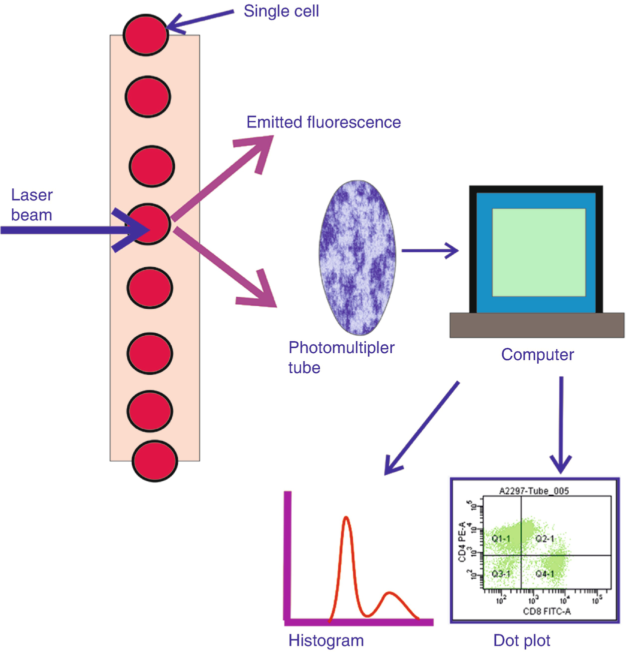
FACScan earlier instruments mandated nylon mesh screening s the smaller flow cells on these machines could easily become clogged by clumped or aggregated cells and often this could. Our gating strategy is similar as previously reported 55. 858 784-8396 flowstaffscrippsedu.
Here are 5 ingredients to consider for your FACS buffer. Flow cytometry was performed on a BD FACScan flowcytometry system. Prepare the following buffer in which to suspend cellular samples prior to cell sorting.
In practice there are differences. Flow cytometry permits the detection of transcription factors within discrete immune cell subsets among a heterogeneous population and provides a sensitive approach to analyzing an immune. Flow cytometry FACS staining protocol Cell surface staining Harvest wash the cells single cell suspension and adjust cell number to a concentration of 1-5x10⁶ cellsml in ice cold FACS.
Flow Cytometry Staining Buffer 1X Flow Cytometry Staining Buffer 1X Catalog Number. Sorting Cells based on Flow Cytometry Data. Flow Cytometry Staining Buffer FACS Buffer This basic FACS Buffer is a buffered saline solution that can be used for immunofluorescence staining protocols antibody and cell dilution.
Flow cytometry histograms are a direct tabulation of the frequencies of measured values in a fixed number of channels or bins. Offers a Range Of Blocking Reagents For Use In Western Blotting Research Applications. Cells that cannot be analyzed immediately for.

Easy Viability Staining For Flow Cytometry Biocompare Com Kit Reagent Review

Flow Cytometric Analysis Of B Cell Development In Bone Marrow And Download Scientific Diagram

Flow Cytometry Facs Protocols Sino Biological

Principle Of Optical Flow Cytometry A As Incident Light Beam Hits Download Scientific Diagram
Flow Cytometry Basic Principles Procedure And Applications In Pathology Springerlink

Optimized Flow Cytometric Protocol For The Detection Of Functional Subsets Of Low Frequency Antigen Specific Cd4 And Cd8 T Cells Sciencedirect
Erb Specific Deletion In Cd11c Cells During Eae A Flow Cytometry Download Scientific Diagram

Flow Cytometry Md Anderson Cancer Center

Gating Strategy Used Throughout The Experiments For Flow Cytometry Download Scientific Diagram

Gating Strategy Used Throughout The Experiments For Flow Cytometry Download Scientific Diagram

Flow Cytometry Basics Flow Cytometry Miltenyi Biotec Technologies Macs Handbook Resources Miltenyi Biotec France

Flow Cytometry Facs Protocols Sino Biological

Optimized Flow Cytometric Protocol For The Detection Of Functional Subsets Of Low Frequency Antigen Specific Cd4 And Cd8 T Cells Sciencedirect

Fundamentals Of Flow Cytometry Aat Bioquest
Fundamentals Of Flow Cytometry Aat Bioquest

Pin On Myalgic Encephalomyelitis Chronic Fatigue Syndrome
Flow Cytometry Basic Principles Procedure And Applications In Pathology Springerlink

How To Identify Problems With Flow Cytometry Panel Design Bad Data Part 2 Cytometry And Antibody Technology

Dri Treg Panel